Uploads by Gillian Air
From CFGparadigms
Jump to navigationJump to searchThis special page shows all uploaded files.
| Date | Name | Thumbnail | Size | Description | Versions |
|---|---|---|---|---|---|
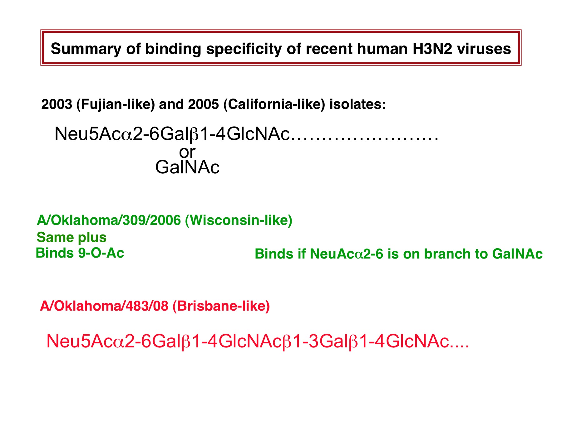
| 03:14, 13 June 2010 | H3 binding specificity.jpg (file) |  |
269 KB | 1 | |
| 03:18, 13 June 2010 | H3 binding.jpg (file) |  |
269 KB | 1 | |
| 04:30, 13 June 2010 | H3binding.png (file) |  |
60 KB | 1 | |
| 05:08, 13 June 2010 | H3binding2.png (file) |  |
24 KB | 1 | |
| 04:40, 4 July 2010 | 5HMG.jpg (file) |  |
285 KB | 1 | |
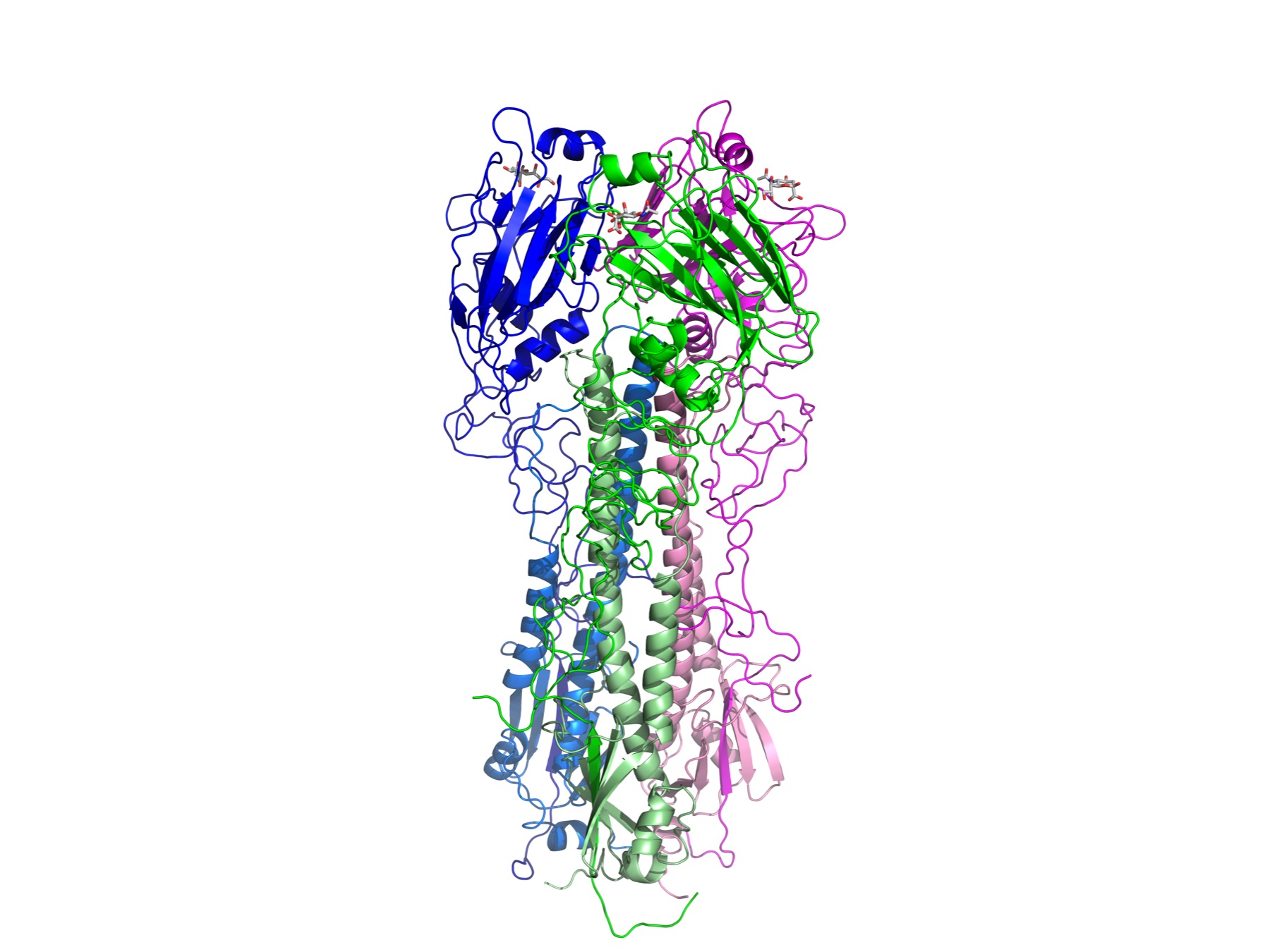
| 04:45, 4 July 2010 | 5HMGjpg.pdf (file) | 288 KB | Structure of the trimer of H3 HA (A/Aichi X-31/68). The image was made using PyMol (Delano Scientific) from PDB file 5HMG. The monomers are colored green, blue and magenta. The darker shade for each is the HA1 polypeptide; the lighter shade is HA2. | 1 | |
| 04:58, 4 July 2010 | 5HMG.png (file) |  |
942 KB | Structure of the H3 trimer. The image was made with PyMol (DeLano Scientific)using PDB file 5HMG. The three monmoers are colored green, blue and magenta.For each, the darker color is the HA1 polypeptide and the lighter shade is HA2. | 1 |