File:5HMG.png
From CFGparadigms
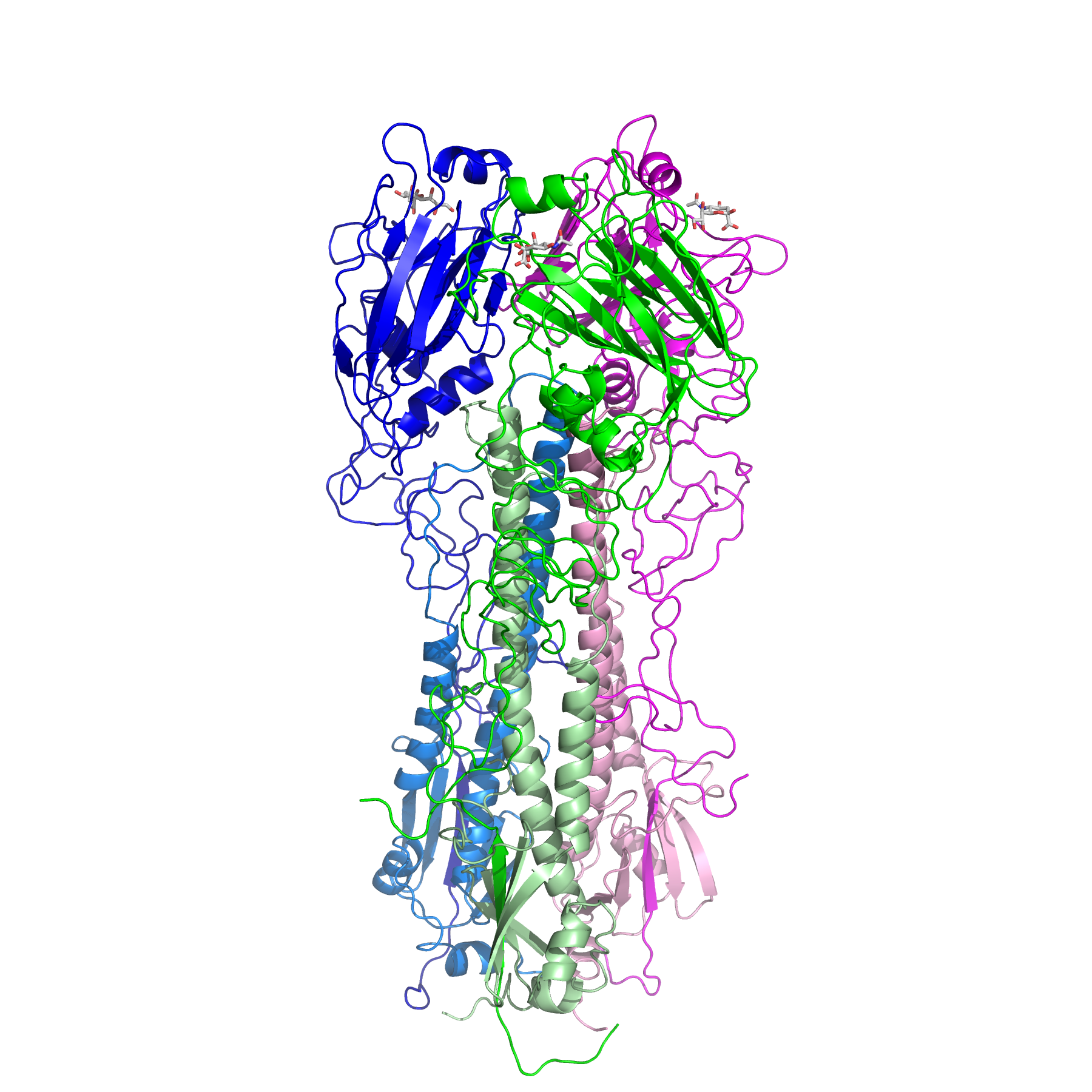
Revision as of 04:58, 4 July 2010 by Gillian Air (talk | contribs) (Structure of the H3 trimer. The image was made with PyMol (DeLano Scientific)using PDB file 5HMG. The three monmoers are colored green, blue and magenta.For each, the darker color is the HA1 polypeptide and the lighter shade is HA2.)
Size of this preview: 600 × 600 pixels. Other resolutions: 240 × 240 pixels | 1,800 × 1,800 pixels.
Original file (1,800 × 1,800 pixels, file size: 942 KB, MIME type: image/png)
Structure of the H3 trimer. The image was made with PyMol (DeLano Scientific)using PDB file 5HMG. The three monmoers are colored green, blue and magenta.For each, the darker color is the HA1 polypeptide and the lighter shade is HA2.
File history
Click on a date/time to view the file as it appeared at that time.
| Date/Time | Thumbnail | Dimensions | User | Comment | |
|---|---|---|---|---|---|
| current | 04:58, 4 July 2010 | | 1,800 × 1,800 (942 KB) | Gillian Air (talk | contribs) | Structure of the H3 trimer. The image was made with PyMol (DeLano Scientific)using PDB file 5HMG. The three monmoers are colored green, blue and magenta.For each, the darker color is the HA1 polypeptide and the lighter shade is HA2. |
You cannot overwrite this file.
File usage
There are no pages that use this file.