Research Highlights
Innate immunity: S. aureus surrenders to mannose-binding lectin
Functional Glycomics (14 February 2008) | doi:10.1038/fg.2008.10Standfirst
Mannose-binding lectin (MBL) binds to Staphylococcus aureus prior to phagocytosis; once engulfed, MBL interacts with Toll-like receptor (TLR) 2 and TLR6 in the phagosome to initiate proinflammatory signaling.
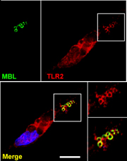
Co-localization (merged image) of MBL and TLR2 on macrophage phagosomes shown by antibody staining. © 2008 The Rockefeller University Press.
Receptors such as Dectin-1 or MBL bind pathogen glycans and act in concert with TLRs as pattern-recognition receptors (PRRs). MBL, a member of the collectin family of lectins, acts as an opsonin to facilitate pathogen phagocytosis, which circumvents the potentially fatal consequences of infections by bacteria such as S. aureus. However, the molecular mechanism by which MBL regulates the immune response to bacterial infection has remained unclear. Now in the Journal of Experimental Medicine, Ip et al. uncover the MBL-dependent pathway that mitigates S. aureus infection in mice.
Serum from wild-type mice infected with S. aureus contained more proinflammatory cytokines and chemokines — including tumour-necrosis factor- (TNF-) and interleukin-6 (IL-6) — than serum from MBL-deficient infected mice. Genetic depletion of the complement protein C3 did not change the MBL-dependent cytokine response, indicating that MBL contributes to a proinflammatory, complement-independent response to S. aureus.
Compared to wild-type mice, S. aureus infection in mice deficient in either TLR2 or MBL resulted in an impaired cytokine response. This result suggests that both PRRs are required for the full immunological response against S. aureus. Furthermore, macrophages that lacked the cytoplasmic TLR adaptor protein MyD88 (myeloid differentiation primary response gene 88) did not react to S. aureus, consistent with the idea that MBL cooperates with TLR2/MyD88 in the same signaling pathway in the response to S. aureus infection.
The authors found that MBL could bind to the bacterial cell wall lipidoglycan lipoteichoic acid (LTA), which is a well-known ligand for TLR2/6. Co-expression of TLR2, TLR6 and MBL in S. aureus-infected human embryonic kidney (HEK) cells activated the TLR effector NF- B, suggesting that S. aureus bacteria induce TLR2/6 heterodimer formation and signaling. Furthermore, MBL and TLR2 were found to form a complex in S. aureus-enriched phagosomes, and cytokine release was strongly diminished when phagocytosis was blocked. These results suggest that MBL and the TLR2/6 heterodimer cooperate to induce TLR signaling following phagocytosis of S. aureus.
Phagosome-associated TLR signaling induced by internalized S. aureus represents a novel mechanism of pathogen defense and stands in contrast to the known immune response to gram-negative bacterial infection, in which bacterial LPS binds to TLR4 on the cell surface. Thus, the results of this study uncover a new means of pathogen defense by glycan recognition, and represent a major advancement in the understanding of S. aureus infection.