Featured Articles
Protein-glycan interaction: An umbrella embraces the virus cone
Functional Glycomics (14 February 2008) | doi:10.1038/fg.2008.7Standfirst
The topology formed by carbohydrate units, and not just their linkage type alone, determines which host cell glycan structures the influenza virus hemagglutinins bind to.
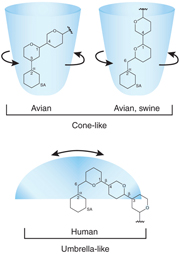
Topologies adopted by animal and human sialoglycans relative to the sialic acid (SA) moiety. From Bewley, C.A.; please click here for a full size picture.
Mutations in the influenza virus can shift the binding specificity of its hemagglutinins from  2-3- to 2,6-linked sialic acids, which terminate epithelial glycans in human upper respiratory tissue. This shift is believed to support the pandemic spread of influenza, such as that of the Spanish flu after the First World War. However, the linkage shift model has been called into question by data that some H1N1 influenza strains — which include the Spanish Flu — can bind to both 2,3- and 2,6-linked sialic acids, even though only a few strains transmit the virus efficiently to humans. Reporting in Nature Biotechnology, Chandrasekaran et al. now clarify this confounding picture.
Chandrasekaran et al. used mass spectrometric analysis of acidic glycans to reveal remarkable diversity in the sialylated glycan receptors in human upper respiratory tissues. The authors found the full spectrum of N-glycan complexity where glycan cores are able to branch into a maximum of four antennae that can be elongated by one or more lactosamine repeats. Although this observation suggests a high diversity of hemagglutinin ligands in the respiratory pathway, examination of glycan- hemagglutinin co-crystal structures revealed that glycans only adopt two main topologies when bound to hemagglutinins. Short N-glycans invariably show a cone-like topology upon hemagglutinin binding regardless of sialic acid linkage. In contrast, long human 2,6-sialylated glycans form an umbrella-like topology (see figure), in which the glycan is bent between the second and third carbohydrate constituent. Chandrasekaran et al. confirmed the suggestion that even distant carbohydrate groups contribute to hemagglutinin binding by showing that the 3rd to 5th carbohydrate residues make numerous contacts with the hemagglutinin molecule. These findings explain the observation that some virus strains bind to short 2,6-sialylated glycans via their hemagglutinins without transmitting the virus efficiently to humans.
The authors screened hemagglutinin-binding glycan array data to verify whether the binding patterns of various hemagglutinins correlate with virus transmitability. Chandrasekaran et al. indeed confirmed that hemagglutinins from human virus strains bind to long 2,6-sialylated glycans, whereas short 2,6- and 2,3-sialylated structures are the sole receptors for avian strains. Furthermore, the author's findings highlight that glycan-hemagglutinin binding is dose-dependent. Avian H5N1 strains were found to bind long 2,6-sialylated glycans when the concentration of the virus particles was very high. As most binding studies are performed without taking into account this dose-dependency, using high virus particle concentrations may cause confounding results about preferred structures.
The authors conclude that the cone or umbrella topology, and not just the linkage type, determines hemagglutinin binding. It appears that, at least in the case of N-glycans, the entirety of the glycan chain delivers structural information that influences binding specificity. Furthermore, the discovery of drugs that bind glycan ligands — such as antiviral medicines — may have to account for a larger complexity of carbohydrate structures.
Raw data from Chandrasekaran et al. is posted on the Functional Glycomics Gateway and can be accessed using this URL