Featured Articles
Dendritic cell receptors: Keeping it in the family
Functional Glycomics (13 March 2008) | doi:10.1038/fg.2008.13Standfirst
The dendritic cell (DC) C-lectin-like immune receptor (DCIR) is closely related to other C-lectin like receptors (CLRs) in its antigen presentation function and inhibitory cross talk with Toll-like receptors (TLRs).
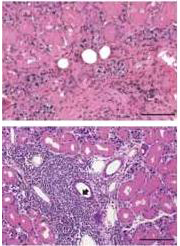
Histopathology of the and salivary gland of 12-month-old wild-type (top) and DCIR-/- mice (bottom; arrow indicates inflammatory changes). Click here for a larger picture. From Fujikado et al.
CLRs recognize viral glycoproteins and cooperate with TLRs in the innate immune response. In contrast to well-characterized CLRs such as DC-SIGN (DC-specific intercellular adhesion molecule 3 grabbing non-integrin), the properties of DCIR are just beginning to become clear. DCIR possesses an intracellular ITIM (immunoreceptor tyrosine-based inhibition motif), which points to an inhibitory function for DCIR signal transduction in DCs via the tyrosin kinase SHP-1. Two studies pinpoint now signaling and in vivo functions of DCIR.
Reporting in Nature Medicine, Fujikado et al. observed that DCIR-knockout (DCIR-/-) mice developed normally in the first months after birth. However, a large proportion of the DCIR-/-mice showed joint deformations, as well as inflammation of salivary glands and tendon bone insertion sites, within one year of age. In contrast to wild-type mice, aged DCIR-/- mice developed IgM autoantibodies and had significantly higher counts of dendritic and CD4+ T cells, suggesting that a lack of DCIR leads to autoimmune reactions. This hypothesis was supported by the strong exacerbation of collagen-induced arthritis observed in DCIR-/- mice and lethally irradiated wild-type mice receiving DCIR-/- instead of normal DCs. The authors found that upon incubation with granulocyte monocyte colony stimulating factor (GM-CSF) DCIR-/- bone marrow-derived DC precursor cells (BMDCs) differentiated more readily into mature DCs than normal BMDCs. Accordingly, GM-CSF signal mediation by STAT5 (signal transducer and activator of transcription 5) transcription factor activation was enhanced in knockout BMDCs compared to wild-type BMDCs exposed to GM-CSF. This finding suggests that STAT5 activation in wild-type BMDCs is dampened by the DCIR via SHP-1 activity. Together, the results of this study show that a lack of DCIR elicits autoimmune reactions.
In Blood, Meyer-Wentrup et al. describe similar functional properties for human plasmacytoid DC (pDC) DCIRs. The authors discovered that almost 80% of pDC DCIR molecules were endocytosed in clathrin-coated vesicles after TLR9 stimulation. Antibody stimulation of DCIR reduced TLR9-mediated cytokine production by 30-40%, which was consistent with the hypothesis that it is the ITIM domain of DCIR that attenuates cytokine signaling through SHP-1 recruitment to TLR9. Thus, the results from both Fujikado et al. and Meyer-Wentrup et al. add evidence to the hypothesis that CLRs such as DC-SIGN and DCIR regulate TLR immune function.
In addition to its inhibitory function towards TLR action, clathrin-coated endocytosis of DCIR suggested that DCIR itself is also involved in antigen presentation. This idea prompted Meyer-Wentrup et al. to link the immunostimulating keyhole limpet hemocyanin protein to an anti-DCIR antibody. Incubation of pDCs with the antigen construct elicited a strong proliferation of T cells, indicating that the content of the clathrin-coated DCIR vesicles is transferred to the pDC antigen presenting pathway, which elicits T-cell proliferation. This finding opens up the possibility of using the DCIR for DC antigen delivery and presentation, and again relates DCIR to DC-SIGN and DEC-205, which are putative vaccine boosters due to their ability to deliver carbohydrate antigens to DCs. Due to the close functional relationship of DCIR to other CLRs it will be interesting to identify the glycan ligand of DCIR's carbohydrate-binding domain.