Research Highlights
Muscular dystrophy: Dystroglycan's unique features
Functional Glycomics (10 July 2008) | doi:10.1038/fg.2008.33Standfirst
An 18-meric  -dystroglycan sequence has been proposed as the preferred sequence for O-mannosylation, and new research shows that sequences upstream of O-mannosylation sites may also guide the mannosyltransferase complex.
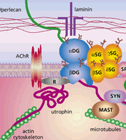
The dystrophin-associated protein complex at the mammalian neuromuscular junction. This complex acts as a receptor for laminin, agrin and perlecan, and functions to link the ECM to the actin cytoskeleton in muscle. From Chamberlain, J. The dynamics of dystroglycan. Nature Genetics 23, 256–258 (1999).
Walker–Warburg syndrome, congenital malformations of brain, muscles and eyes caused by a lack of -dystroglycan O-mannosylation during embryogenesis, is lethal soon after birth. Unique among all glycosylation types known in mammals, the mucin domain of -dystroglycan is the only known target of the mannosyltransferase complex. Identifying the subgroup of serines or threonines that attracts O-mannosylation may reveal how the mannose-based tetrasaccharides guide -dystroglycan's interaction with extracellular laminin. Breloy et al., in the Journal of Biological Chemistry, now provide evidence that complements the mannosylation study of Manya et al. published in the same journal.
Both groups worked with fragments of the human -dystroglycan mucin domain and used the mannosyltransferase complex of the same human cell line. Manya et al. analyzed the O-mannosylation kinetics of native and mutated 20-meric -dystroglycan peptides by in vitro assays. They found that human mannosyltransferases prefer threonines adjacent to prolines in the 18-meric IXPT(P/X)TXPXXXXPTX(T/X)XX sequence occurring twice in the -dystroglycan mucin domain.
Breloy et al. generated fusions of -dystroglycan mucin domain fragments and analyzed the products of the in vivo O-mannosylation by mass spectrometry. In contrast to the in vitro study, Ser/Thr clusters downstream of the proposed consensus sequence, flanked by diads of basic amino acids, were found to be O-mannosylated. The authors point out that the identified -dystroglycan sequences did not exhibit a common sequence motif. Instead, they found that the fusions were O-mannosylated only if a 40-meric sequence located upstream of the glycosylation site was present.
Thus, further research may be required to clarify the substrate preferences of O-mannosylation. Although the complex O-mannosylation sequences identified by both groups seem to be unique to -dystroglycan, other proteins may possess parts of these sequences and therefore also attract O-mannosylation.