Featured Articles
Proteoglycan editing: Sulfatases change a frog's fate
Functional Glycomics (11 September 2008) | doi:10.1038/fg.2008.39Standfirst
Extracellular sulfatases edit cell-surface heparan sulfate proteoglycans, thereby modulating signaling pathways that determine Xenopus tropicalis development.
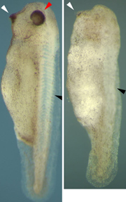
Loss of anterior structures in X. tropicalis embryo upon Xtsulf1 knockdown (right; left: control X. tropicalis embryo). Image by courtesy of Dr Betsy Pownall and Dr Steve D. Freeman, Biology Department, University of York.
Up to 200 variously sulfated disaccharides — N-acetylglucosamine linked to hexuronic acid — form heparan sulfate. It is attached to heparan sulfate proteoglycans (HSPGs), among them growth factor co-receptors such as glypicans and syndecans. Despite the variability in length and degree of sulfation, specific heparan sulfate structures influence cellular functions in different ways. The variability of heparan sulfate derives from enzymes modifying the polysaccharide during its synthesis in the Golgi apparatus. Moreover, tissue-specific sulfatases localized on the cell surface, which preferentially cleave 6O-sulfate groups of trisulfated disaccharides, further enhance the structural complexity of heparan sulfate molecules carried by HSPGs. Two studies now add to our knowledge about the biochemical properties and developmental effects of these cell-surface sulfatases.
Analyzing HSPGs from Sulf knockouts in mouse embryonic fibroblasts, Lamanna et al., writing in the Journal of Biological Chemistry, found that loss of Sulf1 or Sulf2 activities results in differential changes in HSPG sulfation. One example, the glypican HSPG fraction, showed that a Sulf1 knockout only increased 6O-sulfation of trisulfated — but not of mono- or disulfated — disaccharides, whereas a loss of Sulf2 boosted the amount of all 6O-sulfate-containing disaccharides by 50%. Surprisingly, a knockout of Sulf2 also caused a decrease in intracellular 2O- and 6O-sulfotransferase expression, pointing to a feedback mechanism between Sulf2 and the expression of the sulfotransferases.
HSPGs are known to enhance fibroblast growth factor (FGF) signaling, as the proteoglycans promote FGF dimerization and FGF receptor subunit clustering. Lamanna et al. found that the amount of phospho-Erk (extracellular signal regulated kinase), an indicator of FGF signaling strength, increased drastically in the double knockout relative to wild-type mouse embryonic fibroblasts. Mirroring these results, Freeman et al., investigating the role of the Xtsulf1 sulfatase in Xenopus tropicalis development, found an increase in diphospho-Erk using knockdown of Xtsulf1 in the frog embryo. Thus, results from both studies indicate that extracellular sulfatase activity attenuates the enhancement of FGF signaling by HSPGs.
Freeman et al., reporting to Developmental Biology, observed that Xtsulf1 expression is temporally regulated and regionally restricted during X. tropicalis embryonic development. A knockdown of XtSulf1 by XtSulf1 antisense morpholino injection into the zygote led to a loss of anterior structures (see image). This phenotype recapitulates an overactivation of FGF signaling and indicates that the dynamic expression of Xtsulf1 may modulate signaling pathways governing X. tropicalis development.
Indeed, the authors found Xtsulf1 and wnt11 were co-expressed in the X. tropicalis embryo, and injection of both Xtsulf1 and wnt11 mRNA — but not injection of one mRNA — into the four-cell stadium led to a high percentage of duplicated body axes. Thus, sulfatase action may promote canonical Wnt signaling in X. tropicalis. In contrast, the association of bmp4 (bone morphogenetic protein 4) and its receptor was found to be reduced in the presence of Xtsulf1, and the expression of BMP target genes was downregulated when Xtsulf1 was overexpressed. These results suggest that BMP signaling also belongs to the signaling pathways inhibited by XtSulf1 activity.
Thus, extracellular sulfatase action shows again that glycan editing influences ligand–receptor interactions. Future studies may reveal further surprising effects of heparan sulfate fine structure on cellular signaling.