Research Highlights
Lectin microarray: A fast scanner for sweet changes
Functional Glycomics (11 September 2008) | doi:10.1038/fg.2008.42Standfirst
A novel lectin microarray discriminates living cells by their surface glycan markers.
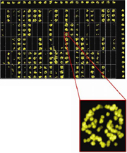
A typical lectin microarray image in a cell binding experiment and an enlarged image of one spot. From Tao, S.-C. et al. Lectin microarrays identify cell-specific and functionally significant cell surface glycan markers. Glycobiology 14 July 2008, reproduction by permission of Oxford University Press.
Changes in cell surface glycans accompany numerous biological processes such as immune-cell maturation. Lectins have been used to track glycosylation changes, and lectin microarrays have been proposed for profiling glycoproteins and glycans in cell lysates. Now, in Glycobiology, Tao et al. present a lectin microarray that detects changes in the cell-surface glycosylation of living cells.
The authors designed a microarray from 94 plant lectins and analyzed the binding of fluorescence-labeled cells. Immature T cells showed a clearly distinguishable binding profile compared with naive and activated T cells, which, in turn, bound to different lectins depending on the activation agent. Interestingly, the lectin binding of activated B cells and activated T cells overlapped, showing that lymphocyte activation elicits similar, but not congruent, glycan-marker expression. In conclusion, the lectin microarray clearly discriminated the maturation stages of lymphocytes.
Cancer stem cells are thought to be the more enduring and aggressive fraction of a cancer cell population, and they may also express specific glycan markers. Based on this hypothesis, Tao et al. analyzed the lectin-binding profile of MCF7 breast cancer cells that were grown conventionally or in sphere culture, by which they are known to express stem-cell-like features. They found that MCF7 cells, but not MCF7 stem-cell-like sphere cells, bound to tomato (Lycopersicon esculentum) lectin (LEL), suggesting that LEL distinguishes both cancer cell populations. Indeed, mice injected with LEL-depleted MCF7 cells developed much larger tumors than mice injected with undepleted MCF7 cells, indicating that depletion by LEL enriches stem-cell-like cancer cells carrying specific glycan surface markers.
Tao et al. used the lectin microarray also to probe bacterial tropism, the preference of bacterial strains to colonize specific tissues. The authors found that the binding strength of various cell lines to mannose-specific lectins mapped the binding of mannose-specific Escherichia coli strains to the cell lines. Thus, the lectin microarray also predicted E. coli tropism. In summary, the lectin microarray affords a fast identification of important cell-surface biomarkers. A further refinement of the technique and an inclusion of animal lectins may increase its potential for high-throughput glycoprofiling.