Featured Articles
Protein O-glycosylation: Drosophila on acid
Functional Glycomics (09 October 2008) | doi:10.1038/fg.2008.44Standfirst
O-glycan profiling of Drosophila melanogaster reveals a wide abundance of glucuronic acid and a concentration gradient of a fucosylated trisaccharide known to modify the Notch receptor.
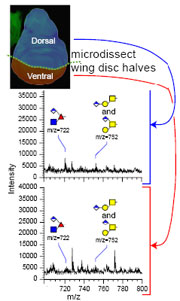
Mass spectrometry shows enrichment of the Notch O-fucose trisaccharide and two further glucuronylated glycans in the dorsal half (top spectrum) of the Drosophila embryo wing disc. © 2008 by the American Society for Biochemistry and Molecular Biology.
Protein O-glycosylation has numerous functions in cell signaling, adhesion and immunology. Apart from mucin-like O-glycosylation and glycosaminoglycosylation, smaller and less frequent fucose- or glucose-based glycans modulate signaling pathways such as for Notch. Although non-vertebrate O-glycomes are believed to be less complex than those of vertebrates, O-glycoprofiling of a model organism such as Drosophila melanogaster has not been performed. Now, in the Journal of Biological Chemistry, Aoki et al. report the O-glycome of the Drosophila embryo.
Mass spectrometry of the O-glycans released from Drosophila embryos showed that the T antigen, a galactose-N-acetylgalactosamine disaccharide also called core 1 O-glycan, was the most abundant O-glycan present. Apart from this structure, small amounts of other, more complex O-glycans — which had not been previously identified in Drosophila — were also detected, including a series of structures branched or terminated with glucuronic acid. One type of a glucuronylated O-glycan had been reported by independent research, yet Aoki et al. found it as a terminating sugar of several hitherto unknown glycan structures. These findings indicate that Drosophila mucin-type O-glycosylation is less complex than in vertebrates, but harbors unusual features not found in mammals.
The authors detected a trisaccharide initiated by O-fucosylation which comprised 20% of all O-glycan structures in the Drosophila embryo. In vertebrates, fucosylation of the Notch receptor by the fucosyltransferase OFUT1 is the first building block of a tetrasaccharide that is required for the binding of Delta to Notch to initiate Notch signaling. Instead of a linear tetrasaccharide, the Drosophila fucose trisaccharide was found to be branched, with glucuronic acid linked to the fucose residue as one of the branches. Interestingly, in mammals, the Notch glycan is terminated by sialic acid which is also an acidic sugar. Glucuronic acid might have similar functions in Drosophila as sialic acid in vertebrates. However, glucuronic acid in Drosophila was also detected within the backbone chain of some glycans, which has never been reported for sialic acid.
To elucidate whether the Drosophila Notch glycan replicates its vertebrate counterpart, Aoki et al. dissected embryo wing discs into dorsal and ventral halves and analyzed the distribution of the Notch trisaccharide. The amount of the fucose glycan in the dorsal half was three times higher than in the ventral half. Importantly, this paralleled the expression of the Fringe glycosyltransferase, which is necessary for elongation of O-fucose, and of the Notch ligand Delta in the dorsal half of the embryo. These results suggest that Drosophila and vertebrate Notch glycans are different in structure yet identical in function.
The results will encourage further research into the role of glycosyltransferases in Drosophila. The characterization of developmental roles for glucuronyltransferases, which might be responsible for modifying O-fucose or core 1 glycans, can now proceed with an understanding of the expected diversity of their products.