Research Highlights
T cell maturation: The ups and downs of sialylation
Functional Glycomics (09 October 2008) | doi:10.1038/fg.2008.46Standfirst
Mice lacking the sialic acid transferase ST6Gal I show a significant reduction in T cells throughout T-cell maturation and an upregulation of proapoptotic genes.
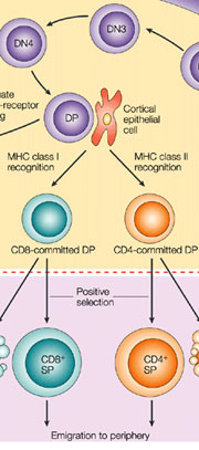
T-cell development in the thymus. Detail from Germain, R. N. T-cell development and the CD4–CD8 lineage decision. Nat Rev Immunol 2, 309–322 (2002). Click here for a full-size picture.
The functional importance of differential sialylation in the immune system has been clarified by previous studies using sialyltransferase-deficient mice.  2,3-bound sialic acid can be transferred by ST3Gal IV and ST3Gal I transferases; however, loss of ST3Gal IV transferase leads to defective lymphocyte rolling, whereas loss of ST3Gal I transferase increases CD8 T-cell apoptosis. When 2,6-linked sialic acid is transferred by ST6Gal I it leads to defects in B-cell function. Now in Glycobiology, Marino et al. report the effects of ST6Gal I deficiency on T-cell development in mice.
Marino et al. used microarrays to analyze gene expression and found that expression of the ST6Gal I transcript varied across different thymocyte developmental populations. ST6Gal I expression increased steeply from the DN3 (double negative 3) to the DN4 T cell stage and in the double positive stage concomitant with intermediate T-cell receptor expression. Levels of ST6Gal I transferase also continued to rise during the transition from double to single positive T cells. The variation in 2,6 sialylation during T-cell maturation was also underscored by lectin binding studies, which indicated that the sialic acid-2,6-galactose glycoepitope was abundant in DN1 and DN4 cells, but very low in the intermittent stages.
The observed changes in ST6Gal I transcript expression prompted Marino et al. to investigate thymocyte populations in ST6Gal I knockout mice (ST6Gal I-/-). Whereas total thymocyte numbers were comparable in both knockout and wild-type mice three weeks after birth, the knockout mice had fewer total thymocytes by 8 and 12 weeks of age. A loss of thymocyte cellularity in ST6Gal I-/- mice was seen in all thymocyte subpolations (DN1-4, double and single positive cells) by 8 weeks of age and was not attributable to an increase in non-T lineage cells. Interestingly, in ST6Gal I-/- mice the transcript abundance of apoptotic genes — such as CD279 in DN1 and CamKIIb in DN2 and DN3 cells — was upregulated when compared to their wild-type counterparts.
While the mechanism leading to the reduction of thymocytes in the ST6Gal I knockout mice has yet to be clarified, it will be interesting to uncover the relationship between the upregulation of apoptosis-related genes and the loss of ST6Gal I.