Research Highlights
O-glycosylation: Messing around with angiogenesis
Functional Glycomics (13 November 2008) | doi:10.1038/fg.2008.49Standfirst
Endothelial cell mucin-type core-1 glycans are required for the separation of blood and lymphatic vasculature in mice.
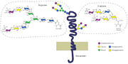
O-glycosylation starts with the addition of a single monosaccharide, such as  -GalNAc in the conventional mucin-type O-glycosylation, attached to serine or threonine.
From van Kooyk & Rabinovich; click here for the original image. © 2008 Nature Publishing Group.
Mice globally lacking mucin-type core-1 O-glycans formed by T-synthase (also known as C1galt1 glycosyltransferase) die around embryonic day 12 from bleeding and failed blood-vessel formation. However, are vascular endothelial cell core-1 O-glycans, which are abundant, required for normal angiogenesis during embryonic development? And do these glycans influence the development of the lymphatic system from lymphatic endothelial cells that, in turn, derive from venous endothelial cells? Reporting to the Journal of Clinical Investigation, Fu et al. analyzed the consequences of a targeted C1galt1 deletion restricted to endothelial and hematopoietic cells (EHC T-syn-/-) to answer these questions.
A backbone of N-acetylgalactosamine galactosylated by C1galt1 forms the base of mucin-type core-1 O-glycans. Fu et al. found that blood and lymphatic endothelial cells of EHC T-syn-/- mice carrying the targeted C1galt1 deletion exposed O-linked N-acetylgalactosamine, thus indicating that the deletion of C1galt1 disrupted galactosylation of N-acetylgalactosamine only in cells forming the vasculature.
Similar to mice globally missing the T-synthase, most EHC T-syn-/- mice died after embryonic day 14.5 or within one week after birth, owing to specific deficits in vascular development. Furthermore, the authors found that lymphatic vessels of EHC T-syn-/- mice were filled with blood, which indicated an insufficient separation of blood and lymphatic vasculature. Hematopoietic cell-specific T–synthase-deficient mice did not reveal these vascular abnormalities, adding to the evidence that a lack of T-synthase in endothelial cell O-glycan formation caused the phenotypic defects. Mice with an induced deletion of T-synthase developed similar vascular defects at six months, indicating that endothelial O-glycans are required for the maintenance of distinct blood and lymphatic vascular systems during postnatal development.
When Fu et al. probed glycoprotein expression by reverse-transcription PCR and western blotting, they discovered that the amount of podoplanin was drastically decreased in EHC T-syn-/- endothelial cells. Importantly, podoplanin-deficient mice phenocopied EHC T-syn-/- mice, suggesting that the podoplanin glycoform synthesized by the T-synthase is required for normal lymphangiogenesis. Cells lacking the enzyme showed strongly reduced surface expression of podoplanin with no intracellular accumulation, indicating that T-synthase expression may be required for podoplanin expression.
Taken together, the results of this study elucidate the function of mucin-type core-1 O-glycans for normal embryonic and postnatal lymphangiogenesis. Targeted deletions of further glycosyltransferases participating in N- or O-glycan formation may increasingly uncover the contributions of specific glycans to embryonic development and postnatal organ growth and tissue repair.